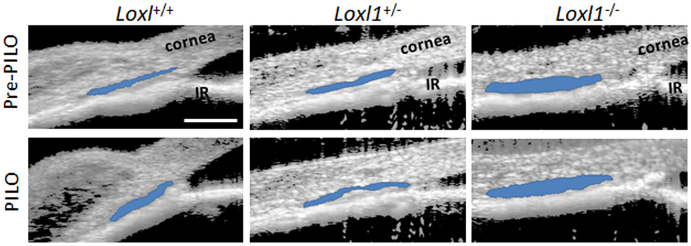
Figure 8. Pilocarpine alters sizes of conventional outflow structures in Loxl1−/− mice.
Sagittal sections of mixed background Loxl1−/−, Loxl1+/− and Loxl1+/+ mouse anterior segments were imaged using SD-OCT at IOP 10 mmHg prior and post topical 1% pilocarpine treatment. SC lumen was segmented using custom software. Data show representative mouse anterior segment OCT images from Loxl1−/−, Loxl1+/− and Loxl1+/+ mice pre- and post-pilocarpine treatment (n=3 for Loxl1+/+, n=4 for Loxl1+/− and Loxl1−/− mice). Segmentation of SC is indicated by blue color. PILO, pilocarpine; IR, iris. Blue area, SC. Scale bar=100μm.

