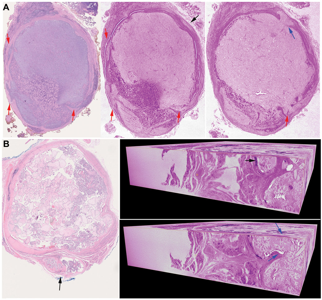
Figure 2. Capsular invasion (CI) demonstrated by using whole slide imaging (WSI) and whole block imaging (WBI) by microCT.
(A) A Hurthle cell carcinoma with multiple foci of CI. Left: WSI of the initial H&E shows 3 foci of CI (red arrows), Middle and right: microCT allows the detection of an additional focus of CI as a satellite nodule (middle, black arrow) on one virtual cut which ends up showing as a mushroom-shaped tumor bud with point of penetration (right, blue arrow) on a different cut. The micro CT images are digitally colored to give an H&E-like appearance. (B) A papillary thyroid carcinoma classic variant with one focus of CI (black arrow) on H&E slide (left). Right: 3D microCT images of the entire paraffin block showing a focus of CI as satellite nodule (black arrow) on one vertical virtual cut which ends up showing as tumor bud penetrating the capsule (point of entry identified by blue arrows) on a different vertical cut.

