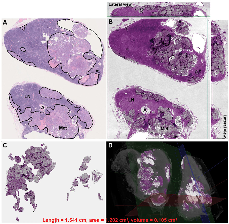
Figure 5. Assessment of metastatic papillary thyroid carcinoma burden in lymph nodes using whole slide imaging (WSI) (A) and whole block imaging (WBI) by microCT (B, C,D).
The area of metastasis is highlighted by black lines and the total surface area was calculated in WSI of H&E (A). Structures, e.g. uninvolved lymph node (LN), adipose tissue (A), and metastasis (Met), are readily identified in both WSI and WBI. The micro CT image in B is digitally colored to give an H&E-like appearance. The total volume of metastasis can be highlighted and calculated in 3D microCT images of WBI. (C) The background uninvolved lymph node is subtracted revealing only the metastatic focus. Note the irregular configuration of the metastatic focus in 3D. (D) The metastatic tumor volume highlighted in bright color in 3D.

