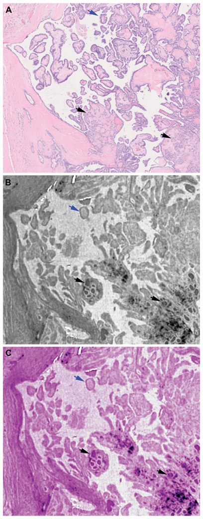
Figure 6. Visualization of papillae and follicles in a papillary thyroid carcinoma, classic using whole slide imaging (WSI) and whole block imaging (WBI) by microCT.
A: WSI of H&E slide with papillae (blue arrow) and follicles (black arrows). B and C: microCT images in greyscale (B) or digitally colored to give an H&E like appearance (C). Papillae (blue arrows) and follicles (black arrows) are easily identified by microCT.

