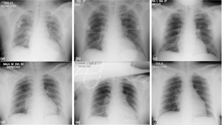
Figure 1.

Serial chest radiographs showed significant recovery in 2 COVID‐19 patients after treatment with itolizumab. Patient 1: (a) (Before itolizumab): bilateral hilar vascular thickening, greater on the right side. Right para‐cardiac shadow opacity. (b) (48 h after itolizumab): decreased bilateral hilar vascular thickening. Decreased opacity in the right para‐cardiac shadow and thickening of the right basal hilum. No pleuro‐pulmonary lesions. (c) (5 days after itolizumab): favorable radiological evolution, with disappearance of bilateral hilar vascular thickening and the right para‐cardiac opacity. No pleuro‐pulmonary lesions. Patient 2: (d) (Before itolizumab): diffuse veil opacities that project in the right para‐cardiac region and in the lower left lung field. (e) (48 h after itolizumab): decrease in veil opacities. (f) (8 days after itolizumab): no pleuro‐pulmonary lesions. Favorable radiological evolution, with disappearance of the lung lesions.
