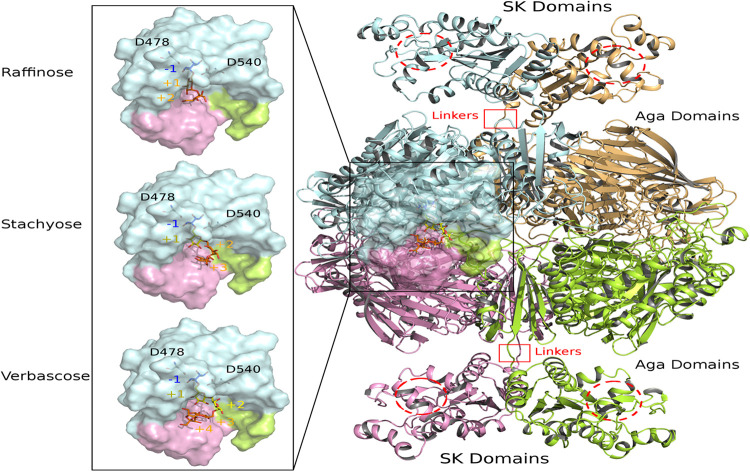
FIGURE 9.
View of modeled quaternary assembly of RgAgaSK-RFO complexes. The tetrameric RgAgaSK structure is shown as carton on the right. Each subunit is highlighted according to a specific color (pale cyan, bright orange, lemon, and pink). SK Domains are located on the top and the bottom of the complex with putative active sites indicated with red dashed circles. Aga domains are viewed as an assembly in the middle, connected to the SK domains by linkers highlighted by red squares. The active site of one Aga subunit is shown as transparent surface. A zoom on this region is displayed on the left with three complexed ligands (raffinose, stachyose, and verbascose) docked independently in Aga active site. For reference, the catalytic residues are shown in ball & sticks at subsite –1. The galactosyl bound in the subsite –1 is colored in blue whereas the sucrose moiety at the end chain is colored in orange. Additional galactosyl units within stachyose and verbascose are colored in yellow.

