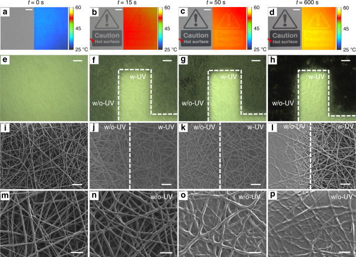
Fig. 2. Pattern activation and changing morphology.
a Photographs (left) and thermal images (right) of fibrous samples after the second step of photo-programming. b–d Photographs and thermal images of the sample in a captured at different times during heating at 55 °C. The color scale in thermal images indicates the temperature of the sample. (Scale bars in (a–d): 5 mm). e–h Corresponding optical micrographs of a–d for a feature of the letter “H” in the sign “Hot” (red arrow in (b–d)). w-UV (w/o-UV) indicates the area of the sample exposed (unexposed) to UV light during the second step of photo-programming. Scale bars, 200 μm. i–l Corresponding SEM micrographs at the interface between the UV exposed (w-UV) and the unexposed areas (w/o-UV), collected at the various time points. Scale bars, 20 μm. m–p Higher magnification view of the areas shown in (i–l), that are not exposed to UV light during the second step of photo-programming. Scale bars, 10 μm.

