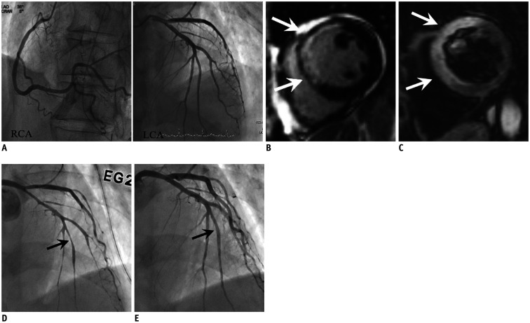
Fig. 2. AMI due to CS in 74-year-old male patient with cardiac arrest after chest pain, elevated troponin (TnI: 4.54 ng/mL) and ECG change (ST elevation in V1–4).
A. Invasive angiograms (RCA and LCA) show no evidence of obstructive coronary artery although increased troponin and ECG abnormality. B, C. Cardiac MR images, which performed due to continuous chest pain, show enhancement at LAD territory (transmural extent 75%) on late gadolinium enhancement (arrows) (B) and hyperintense edema at same territory on T2-weighted image (arrows) (C), indicating AMI of LAD territory. D, E. Thus, invasive coronary angiograms were performed again with ergonovine provocation test and revealed significant stenosis at distal LAD (arrow) (D), which relieved after infusion of nitroglycerin (arrow) (E). AMI = acute myocardial infarction, ECG = electrocardiogram, LAD = left anterior descendingartery, LCA = left coronary artery, RCA = right coronary artery

