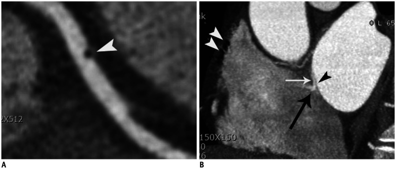
Fig. 4. Coronary thromboembolism in 43-year-old male patient with atypical chest pain who was confirmed as coronary embolism.
A. Curved multiplanar reformatted CT image showed air bubble (arrowhead) in middle RCA. B. Air bubble passed through patent foramen ovale (space between septum primum [black arrowhead] and secondary septum [white arrow]) with contrast jet flow (black arrow). Note that multiple air-bubbles (white arrowheads) are demonstrated in right atrium. Air-bubbles are introduced into right atrium when contrast materials were flushed into right antecubital vein.

