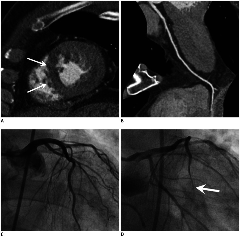
Fig. 5. CS in 65-year-old male patient with acute chest pain and elevated troponin.
A, B. Cardiac CT with short axis view of LV in systolic phase shows hypokinesia and subendocardial hypoperfusion at apical septal wall of LV myocardium (arrows). However, curved multiplanar reformatted image of LAD artery (B) shows no significant stenosis. C, D. Invasive angiograms initially demonstrated no obstructive stenosis. However, the significant stenosis is noted at distal LAD (arrow) (D) after ergonovine provocation test, confirming the CS. LV = left ventricle

