
Figure 1.
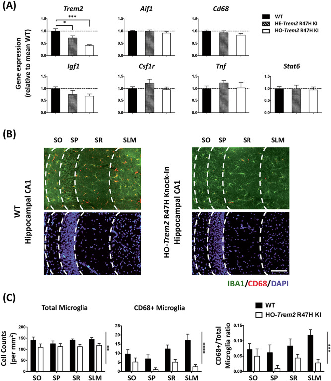
Trem2 R47H KI mice (4 months old) showed significant down-regulation of Trem2 expression, and decreased microglia density and CD68 levels in the hippocampus. (A) Gene expression in hippocampal homogenates from homozygous (HO) and heterozygous (HE) Trem2 R47H KI mice relative to Rps28 and then expressed relative to WT within the batch. N = 6–7 mice per group. Data shown as mean ± SEM. One-way ANOVA showed a significant main effect of genotype for Trem2 expression only, and so Sidak’s post hoc tests were used to test pairwise significance between the three genotypes; *P < 0.05, **P < 0.01, ***P < 0.001. (B) Representative images showing AIF1/IBA1 and CD68 immuno-staining of microglia with DAPI in the hippocampal CA1 regions from WT and HO-Trem2 R47H KI mice. The layers from left are SO (stratum oriens), SP (stratum pyramidale), SR (stratum radiatum), and SLM (stratum lacunosum-moleculare). Scale bar: 100 μm. (C) Left: total microglia density in all four CA1 layers was decreased in HO-Trem2 R47H KI mice. Middle: density of CD68 positive microglia decreased in HO-Trem2 R47H KI mice. Right: proportion of CD68 positive microglia was significantly lower in HO-Trem2 R47H KI mice. N = 5–6 mice per group. Data shown as mean ± SEM. Two-way ANOVA with significant main effect of Trem2 genotype indicated with vertical lines, no significant interactions were seen between hippocampal layer and Trem2 genotype; *P < 0.05, **P < 0.01, ***P < 0.001, ****P < 0.0001.