
Figure 9.
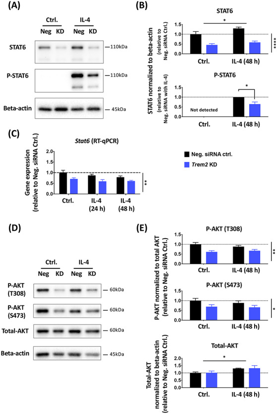
Trem2 knockdown resulted in significantly decreased total STAT6 levels and AKT phosphorylation in primary microglia in vitro. (A and B) Total and phosphorylated STAT6 protein levels in primary microglia treated with IL-4 and Trem2 siRNA were analyzed with western blot. N = 4 independent experiments. (C) Stat6 gene expression levels in primary microglia, assessed by RT-qPCR (N = 3 independent experiments). (D and E) Total and phosphorylated (at Thr308 and Ser473) AKT protein levels analyzed by western blot. N = 4 independent experiments. Protein levels were normalized to beta-actin and gene expression was normalized to Rps28. Fold change was calculated relative to the negative control without IL-4 treatment in each individual culture preparation. Note that phosphorylated STAT6 was not detectable under control conditions and so under IL-4 conditions the effect of Trem2 knockdown was calculated as fold change relative to control cells. Data were shown as mean ± SEM. With the exception of phosphorylated STAT6, data were analyzed by two-way ANOVA; significant main effects of IL-4 treatment and Trem2-knockdown indicated by horizontal and vertical lines respectively, no significant interactions were seen between IL-4 treatment and Trem2 knockdown. Phosphorylated STAT6 was analyzed by a one-sample t-test. *P < 0.05, **P < 0.01, ***P < 0.001, ****P < 0.0001.