
Figure 3.
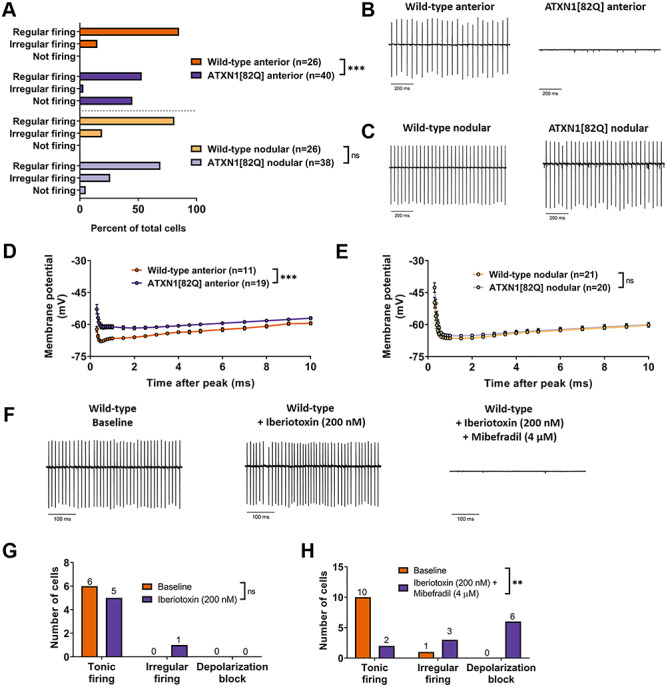
Regionally dysregulated ion channel genes form a functional module critical for Purkinje neuron pacemaking. (A) The distribution of regularly firing, irregularly firing and non-firing cells was recorded for Purkinje neurons in the anterior cerebellum and nodular zone. (B) Representative trace from a tonic firing wild-type and non-firing ATXN1[82Q] Purkinje neuron in the anterior cerebellum at P35. (C) Representative trace from wild-type and ATXN1[82Q] Purkinje neurons in the nodular zone at P35. (D and E) Membrane potential measurements during the AHP in wild-type Purkinje neurons (D) and ATXN1[82Q] Purkinje neurons (E) in the anterior cerebellum at P35. (F) Representative traces of wild-type Purkinje neurons in the anterior cerebellum at P35. Traces are shown at baseline (left), after perfusion of 200 nM iberiotoxin (middle) and after perfusion of 200 nM iberiotoxin +4 μM mibefradil. (G and H) Summary distribution of regularly firing, irregularly firing and non-firing Purkinje neurons before and after perfusion of 200 nM iberiotoxin (G) and after 200 nM iberiotoxin +4 μM mibefradil (H). **Denotes P < 0.01; ***denotes P < 0.001; ns denotes P > 0.05; Chi-square test (A, G and H); two-way repeated measures ANOVA with Holm-Sidak correction for multiple comparisons (D and E).