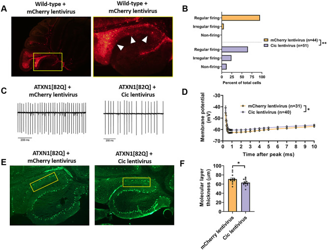
Figure 5.

Increased Cic expression results in Purkinje neuron hyperexcitability and accelerates Purkinje neuron degeneration. (A) Stereotaxic injection of a lentiviral construct containing mCherry results in widespread expression in lobule IX, but not in lobule X, 10 days after injection. (B) The distribution of regularly firing, irregularly firing and non-firing Purkinje neurons in the nodular zone of ATXN1[82Q] mice at 10 days post-injection with Cic lentivirus or mCherry lentivirus. (C) Representative traces of ATXN1[82Q] Purkinje neuron spiking in the nodular zone at 10 days post-injection with Cic lentivirus or mCherry lentivirus. (D) Quantification of the AHP from ATXN1[82Q] Purkinje neurons in the nodular zone at 10 days post-injection with Cic lentivirus or mCherry lentivirus. (E) Representative images of the nodular zone of ATXN1[82Q] mice at 70 days post-injection with Cic lentivirus or mCherry lentivirus. Yellow box indicates analyzed area of lobule IX. (F) Quantification of molecular layer thickness measurements as illustrated in panel (E). *Denotes P < 0.05; **denotes P < 0.01; Chi square test (B); two-tailed Student’s t-test (F).
