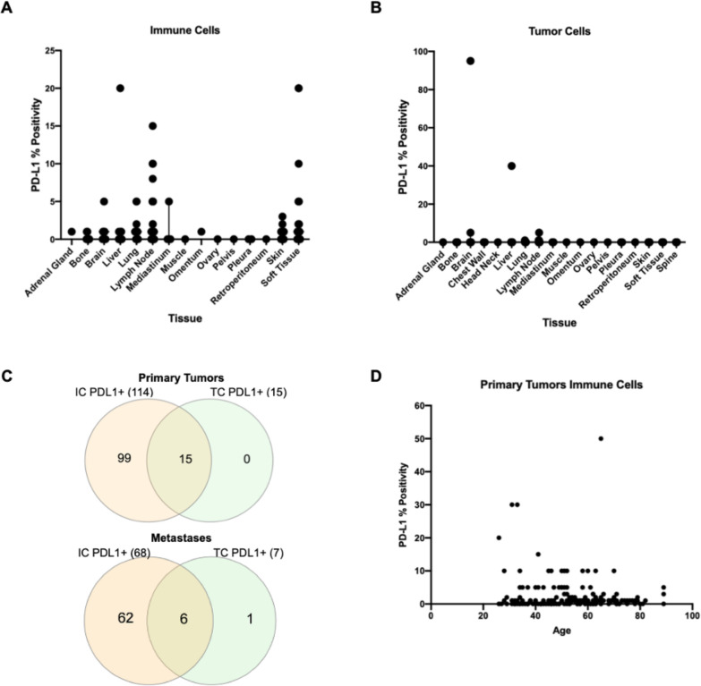
Figure 1.
Programmed Death Ligand 1 (PD-L1) expression. (A) PD-L1 percent positivity by IHC on s immune cells by metastatic location. (B) PD-L1 percent positivity by IHC on tumor cells by metastatic location. (C) Venn diagrams of PD-L1 positive ICs (left) and TCs (right) in breast primaries (N=179) and metastatic lesions (N=161). (D) PD-L1 percent positivity on ICs in primary tumors by age (r=0.02, p value=0.7833). IC, immune cells; IHC, immunohistochemistry; TCs, tumor cells.

