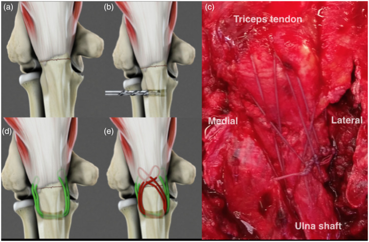
Figure 2.
(a)–(e). Depiction of suture fixation technique. The fracture is reduced (a) and a transverse drill hole is made in the ulna (b). Sutures are passed from lateral to medial with the first set grasping the proximal fragment in a longitudinal manner (c) and the second set creating a crisscross configuration (d). Intra-operative view of the technique used to fix a chevron olecranon osteotomy (e).

