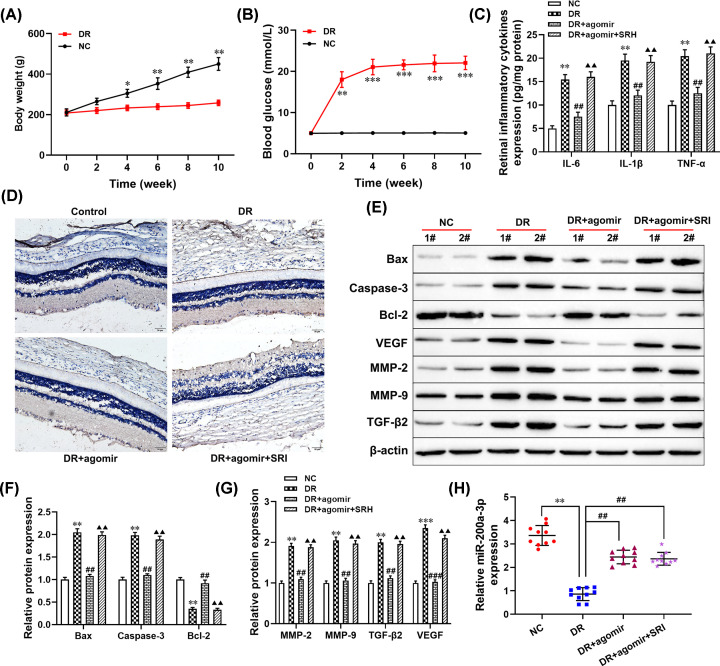
Figure 5. Overexpression of miR-200a-3p attenuated the progression of DR through regulating the TGF-β2/Smad pathway in vivo.
DR model group, all rats received an intraperitoneal injection of STZ (60 mg/kg) which was dissolved in freshly prepared sodium citrate buffer (0.1 mM, pH 4.5); DR+miR-200a-3p group, given agomir (miR-200a-3p; 3 mg/kg) through tail intravenous injection and then received an intraperitoneal injection of STZ; DR+miR-200a-3p+SRI group, all rats were given SRI (1 mg/kg) through tail intravenous injection and then received an intraperitoneal injection of STZ. (A) The body weight of all mice was measured, *P<0.05, **P<0.01, compared with the NC group. (B) The level of blood glucose in all mice was examined, **P<0.01, ***P<0.001, compared with the NC group. (C) The levels of inflammatory cytokines were detected using ELISA. (D) Immunohistochemical staining was performed to detect the expression of VEGF. (E–G) The protein levels of Bax, caspase-3, bcl-2, MMP-2, MMP-9, TGF-β2, and VEGF were measured using Western blotting. (H) RT-qPCR was used to detect the expression of miR-200a-3p. **P<0.01, compared with the NC group; ##P<0.01, compared with the DR group; ▲▲P<0.01, compared with the HG+agomir group.

