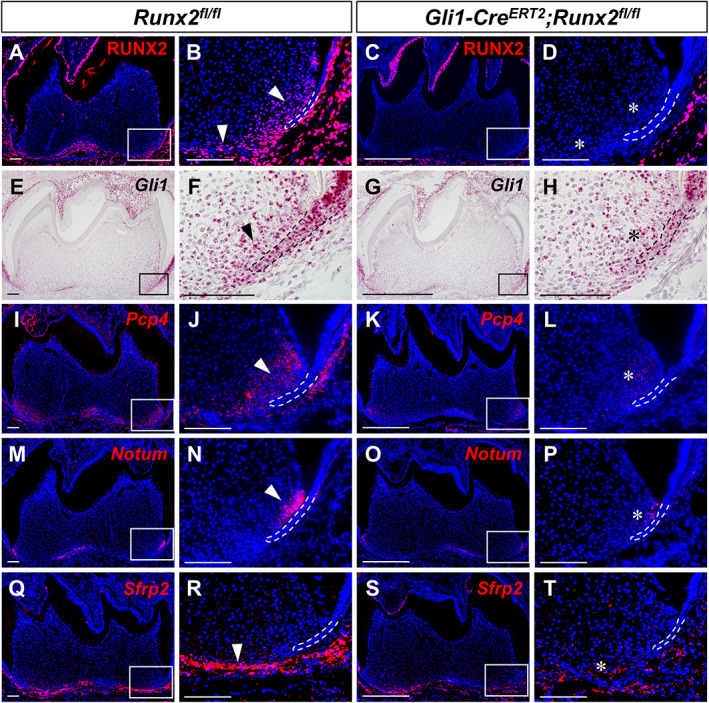
Fig 4.

In vivo validation of putative downstream targets upon deletion of Runx2 in the dental mesenchyme. RUNX2 immunofluorescence (A–D) and RNAscope in situ hybridization (red) of Gli1 (E–H), Pcp4 (I–L), NOTUM (M–P), and Sfrp2 (Q–T) of sagittal sections of mandibular molars from PN7.5 Runx2 fl/fl control and Gli1‐Cre ERT2 ;Runx2 fl/fl mice. The boxed area is enlarged on the right. Dashed lines outline HERS. Arrowhead indicates positive signals; asterisks indicate altered staining in targeted region of mutant samples. n = 3 sections were examined from multiple littermate mice per group. Scale bars = 100 μm.
