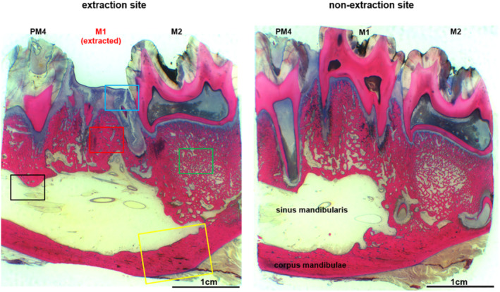
Fig 8.

Histological overview of the region of the left‐sided extraction alveoli M1 of the tooth‐only control group (left image section) compared to the corresponding region of the opposite side (right). There is an intact epithelial integrity, no exposed bone, no signs of inflammation, and no endosteal or periosteal bone proliferation.
