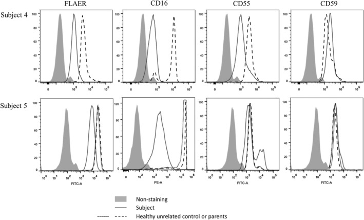
FIGURE 2.

Impact of the PIGQ variants on the expression of GPI‐APs. Flow cytometry analysis of granulocytes from fresh blood from subject 4 (top row) and subject 5 (bottom row) and compared to healthy controls and parents, respectively. Fluorescently labeled proaerolysin “FLAER” directly binds the GPI‐anchor, whereas CD16, CD55, and CD59 all stain for GPI‐APs
