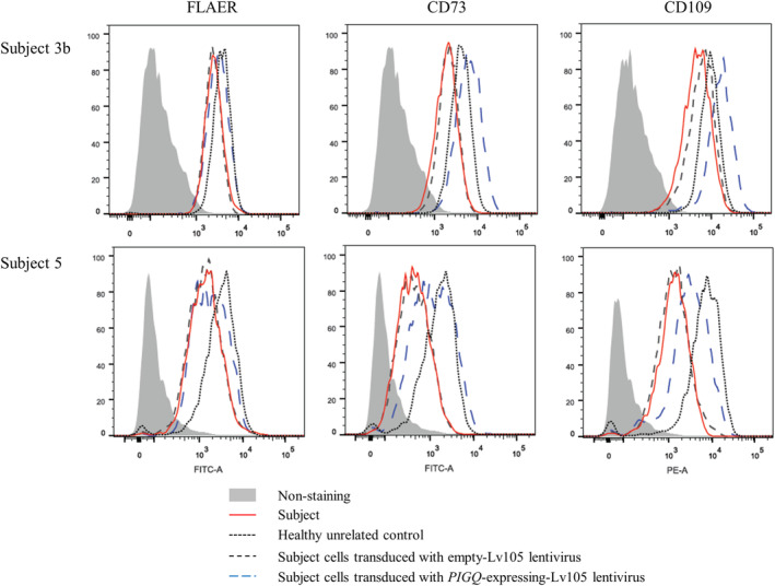
FIGURE 3.

Impact of the PIGQ variants on the expression of GPI‐APs. Flow cytometry analysis of fibroblasts derived from subject 3b (top row) and subject 5 (bottom row) and compared to healthy controls and parental derived cell‐lines, respectively. Subject cell lines were further transfected with empty lentivirus or lentivirus expressing WT PIGQ cDNA. Fluorescently labeled proaerolysin “FLAER” directly binds the GPI‐anchor, whereas CD73 and CD109 stain for GPI‐APs
