
FIGURE 2.
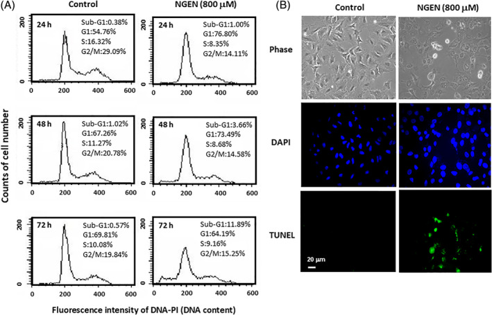
NGEN induces DNA fragmentation as detected by flow cytometry and TUNEL assay. A, The cells were treated with 800 μM NGEN for 24, 48, and 72 hours, and were harvested for flow cytometry analysis of the sub‐G1 population. Quantification of sub‐G1 phase and cell population in each phase was determined using ModFITLT 2.0 software. B, A549 cells were treated without or with 800 μM of NGEN for 48 hours, and then TUNEL assay was performed, and nuclear DNA was stained using DAPI. The stained cells were examined by a fluorescence microscope with a magnification of ×200 (scale bar, 20 μm) [Color figure can be viewed at wileyonlinelibrary.com]