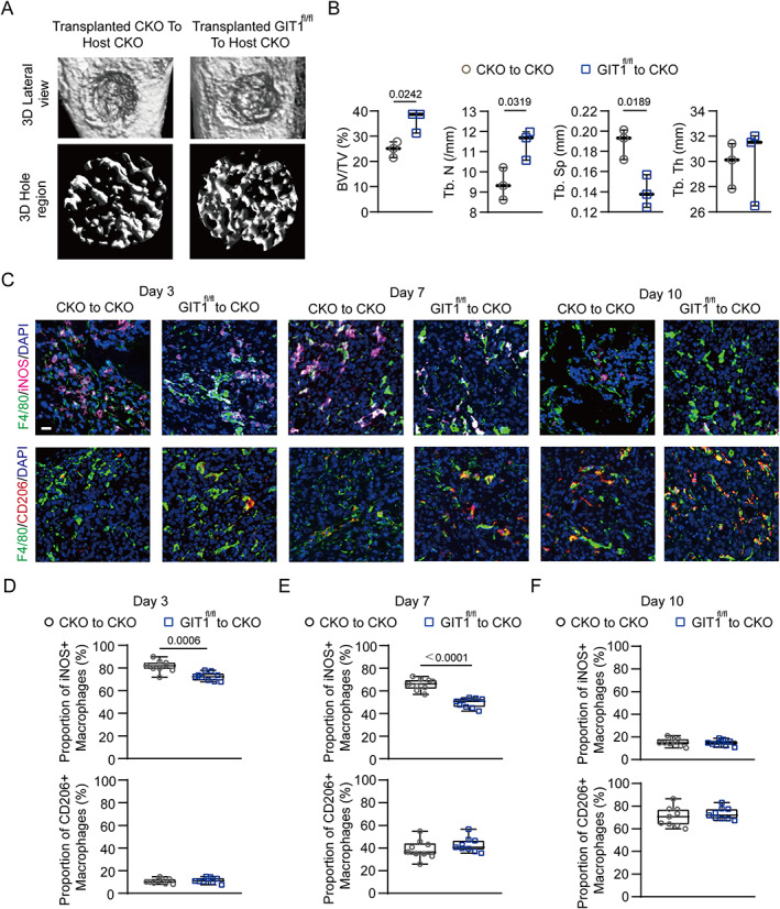
Fig. 7.

Substitution of GIT1 CKO bone marrow with GIT1fl/fl bone marrow facilitates intramembranous bone healing. (A) Micro‐CT reconstruction of tibial defect (top panel) and mineralized bone in the hole region (lower panel) of indicated groups. (B) Quantitative analysis of BV/TV (%), Tb.N, Tb.Sp, and Tb.Th of the regenerated bone in the tibial defect from different groups (unpaired two‐tailed Student's t test). (C) Infiltration of M1‐like (F4/80+ and iNOS+) (top panel) and M2‐like (F4/80+ and CD206+) (lower panel) macrophages in the tibial defect region from indicated groups on days 3, 7, and 10 post‐injury were identified using IF staining. Nuclei were counterstained with DAPI (blue). Scale bar = 100 μm. (D–F) Proportion of infiltrated M1‐like (F4/80+ and iNOS+) and M2‐like (F4/80+ and CD206+) macrophages at indicated time points post‐injury from different transplanted mice (CKO to CKO versus GIT1fl/fl to CKO) were determined (unpaired two‐tailed Student's t test).
