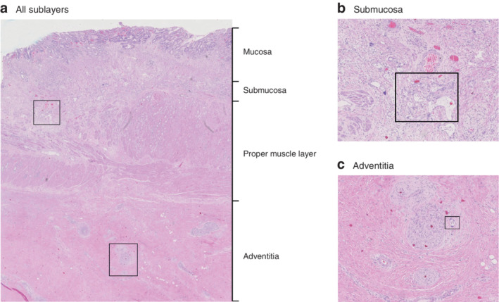
Fig. 1.

Histology of oesophageal resection specimen a Section from an oesophageal resection specimen showing sublayers. Detailed examples of boxed areas in the submucosa and adventitia are shown in b and c respectively. b The boxed area indicates glandular adenocarcinoma within an area of regressional changes in the submucosa. The submucosa was scored as tumour regression grade (TRG) 3 (more than 10 per cent vital tumour cells). c The boxed area shows vital tumour cells within an area of regressional changes in the adventitia. The adventitia was scored TRG 2 (10 per cent or less vital tumour cells). (Haematoxylin and eosin staining; a × 10 magnification, b,c × 40 magnification.)
