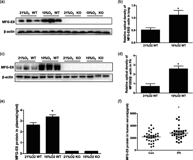
Figure 2.

MFG‐E8 protein expression in chronic hypoxia‐induced mice and PH patients. Representative western blot images of MFG‐E8 in lung (a) and average data (b). Representative western blots in pulmonary arteries (c) and average data (d), n = 3. (e) ELISA detected the MFG‐E8 protein in mouse blood plasma (ng/ml); n = 6; *p < .05 compared with the 21% O2 WT group. (f) MFG‐E8 protein in human blood serum (pg/ml); n = 30 or 35, results are expressed as the mean ± SEM, and *p < .05 compared with the control group. ELISA, enzyme‐linked immunosorbent assay; KO, knockout; MFG‐E8, milk fat globule‐EGF factor 8; PH, pulmonary hypertension; WT, wild type
