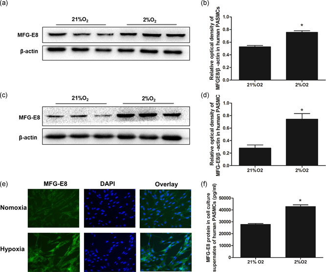
Figure 3.

MFG‐E8 protein expression in human PASMCs. Representative western blot images of MFG‐E8 (a) and average data (b) after 48 hr of exposure to 2% O2. Representative western blots of MFG‐E8 (c) and average data (d) after 72 hr of exposure to 2% O2; *p < .05 compared with 21% O2. (e) Qualitative immunofluorescence staining for MFG‐E8 (green) and nuclei (DAPI) after 48 hr of exposure to 2% O2. (f) ELISAs detected MFG‐E8 protein in collected human PASMC cultured supernatants (pg/ml); n = 3, results are expressed as the mean ± SEM, and *p < .05 compared with the 21% O2 group. DAPI, 4′,6‐diamidino‐2‐phenylindole; ELISA, enzyme‐linked immunosorbent assay; MFG‐E8, milk fat globule‐EGF factor 8; PASMC, pulmonary artery smooth muscle cell
