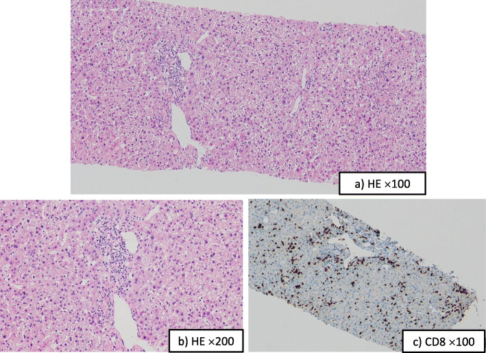
Fig. 3.
Histological findings of the liver. a and b Photomicrographs of a representative hematoxylin and eosin (H&E) section from the liver biopsy (a: H&E, original magnification × 100; b: H&E, original magnification × 200). Moderate inflammation and focal necrosis were observed. The liver parenchyma was mainly damaged with moderate infiltration of lymphocytes. c Photomicrograph shows CD8 immunohistochemistry of a f3:4 representative section from the liver biopsy. Most of the infiltrating lymphocytes were positive for CD8 staining (brown)

