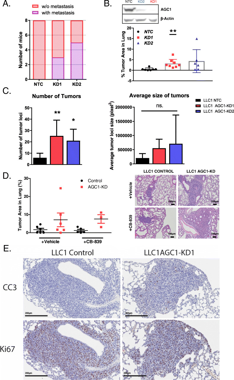
Fig. 2.
Lewis lung carcinoma (LLC1) cells with AGC1-knockdown have higher potential to metastasize. a The number of mice with (purple) or without (pink) lung metastasis 16 days after control (NTC) or AGC1-KD LLC1 cells (KD1, sh911 or KD2, sh908) were injected in the flanks. H&E-stained lung slides were screened for metastasis. b Percent of the metastatic area in the lungs of mice bearing control (NTC), or AGC1-KD (KD1 with sh911; KD2 with sh908) LLC1 tumors, measured 21 days after cells were injected subcutaneously behind the necks of mice (n ≥ 6). Lungs were resected, and H&E-stained sections were analyzed. c (left) Number of metastatic tumor loci and (right) average tumor locus size in the lungs of mice bearing control (NTC), or AGC1-KD (KD1 with sh911; KD2 with sh908) LLC1 tumors, measured 21 days after cells were injected subcutaneously behind the necks of mice (n ≥ 6). Lungs were resected and H&E-stained sections were analyzed. d (left) Percent of the metastatic area in the lungs of mice that were bearing control (NTC), or AGC1-KD LLC1 tumors on the flanks 22 days after injections. Mice were treated without (vehicle) or with CB-839 dosed at 200 mg/kg twice daily starting on day 13 (n ≥ 6). (right) Representative histology pictures of the metastatic areas of mice injected with control (NTC) or AGC1-KD LLC1 tumors. e Representative images of the metastatic areas of vehicle-treated mice (as in d) injected with control (NTC) or AGC1-KD LLC1 tumors, stained with proliferation (Ki67) and apoptosis (cleaved caspase 3; CC3) markers. Images were taken at × 20 magnification. All experiments denote mean ± SDs. Significance levels: * p ≤ 0.05, ** p ≤ 0.01, *** p ≤ 0.001

