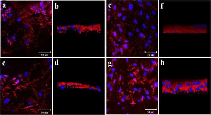
Fig. 4.
Immunofluorescent staining of Runx-2 and fibronectin expression in osteoblasts. Stained with reds are Runx-2 and fibronectin. TPS-coated surface stained with fibronectin is showed in a and b while fibronectin staining of DMF surface is showed in c and d. Runx-2 expression of TPS surfaces is showed in e and f while that of DMF surface are showed in g and h. Blue stains are of DAPI, which were used as counterstain. Overall expression within the set area is shown in a, c, e, and g. The thickness of the stained layer is shown in b, d, f, and h

