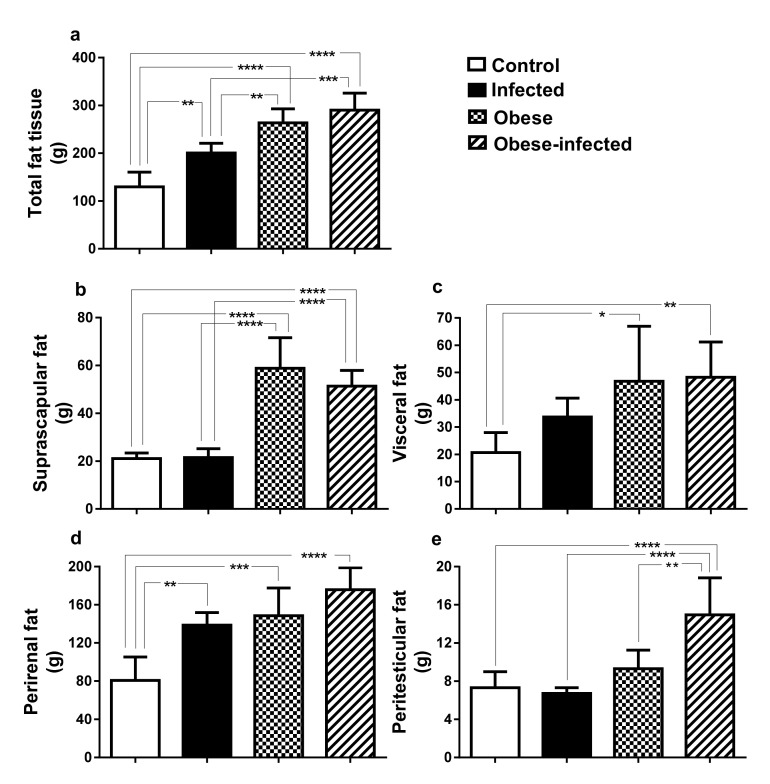
Figure 5.
Distribution of body adipose tissue in rabbits infected with T. pisiformis. (a) weight of total adipose tissue, (b) suprascapular, (c) visceral, (d) perirenal, and (e) peritesticular. Data show mean ± SD. * P ≤ 0.05, ** P ≤ 0.01, *** P ≤ 0.001, **** P ≤ 0.0001, ANOVA test, followed by a Tukey´s post hoc test.

