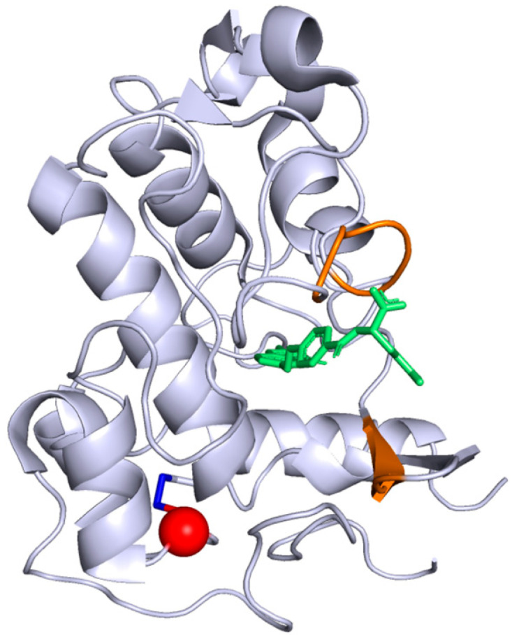
Figure 4.
Crystallographic structure of FRα protein, from the Protein Data Band (4LRH). The folate is in green, the folate binding site is colored in orange. The Cys66Tyr substitution position induced by the pathogenic variant described in our case report is represented in red while the disulfide bond between Cys66 and Cys109 is in dark blue.

