
Figure 2.
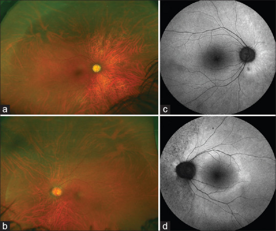
Ultra-wide field fundus imaging of a patient with autoimmune retinopathy (non-paraneoplastic subtype) in a 45-year-old female (a and b). The fundus does not show any significant clinically visible changes. The fundus autofluorescence images (c and d) show very subtle changes especially in the nasal part of the left eye (d). There are stippled areas of hypo-autofluorescence corresponding to the retinal pigment epithelial damage