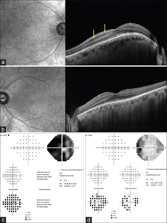
Figure 3.

Spectral-domain optical coherence tomography (SD-OCT) of the same patient shows presence of intraretinal cystic spaces in the right eye (yellow arrows) in the perifoveal region. There is increase in the overall central macular thickness (a). The left eye shows mild increase in central retinal thickness (b). The visual field analysis (c and d) shows severe generalized field loss especially in the right eye and loss of nasal field in the left eye
