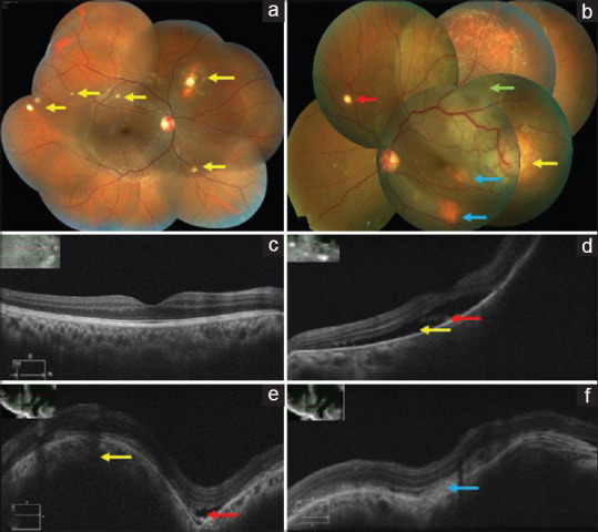
Figure 1.

(a) Right eye color fundus montage photo showing multiple choroidal tubercles (yellow arrows). (b) Left eye color fundus montage photo showing a tubercular granuloma temporal to the macula two-disc diameter in size (yellow arrow) with superior exudative retinal detachment (green arrow) and SRF extending till the macula. Two smaller granulomas noted inferotemporal to the macula and in the inferior quadrant (blue arrows). A single choroidal tubercle noted in the superonasal quadrant (red arrow). (c) Right eye OCT scan showing normal foveal contour. (d) Left eye OCT macular scan showing SRF (yellow arrow) with shaggy photoreceptors (red arrow). (e and f) Left eye OCT scan through the granuloma temporal to the macula showing an elevation of the retinal layers with an underlying choroidal bump (yellow arrow) and a small pocket of SRF (red arrow) and subretinal fibrin (blue arrow)
