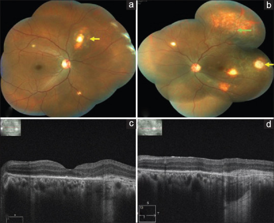
Figure 4.

(a) Right eye color fundus montage photo showing regressed choroidal tubercles (yellow arrows). (b) Left eye color fundus montage photo 1 month after the third injection showing complete regression of the granuloma (yellow arrow) with scarring and resolved exudative retinal detachment (green arrow). (c) Left eye OCT macular scan showing complete resorption of SRF. (d) Left eye OCT scan through the granuloma showing flattening of the RPE with complete resolution of SRF
