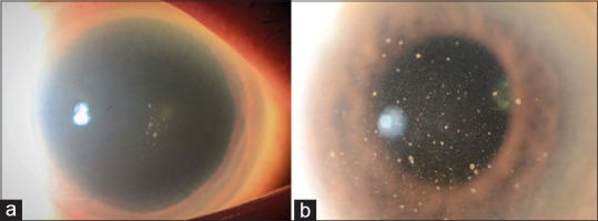
Figure 3.

Slit-lamp photograph of CMV AU in diffuse illumination showing a couple of granulomatous keratic precipitates in the center of the steamy cornea in an eye presenting acutely with elevated intraocular pressure typical of CMV associated Posner Schlossman syndrome (a) and diffusely distributed fine filiform keratic precipitates typical of Fuchs uveitis syndrome admixed with scattered pigmented, medium-sized keratic precipitates in an eye with cytomegalovirus chronic anterior uveitis (b)
