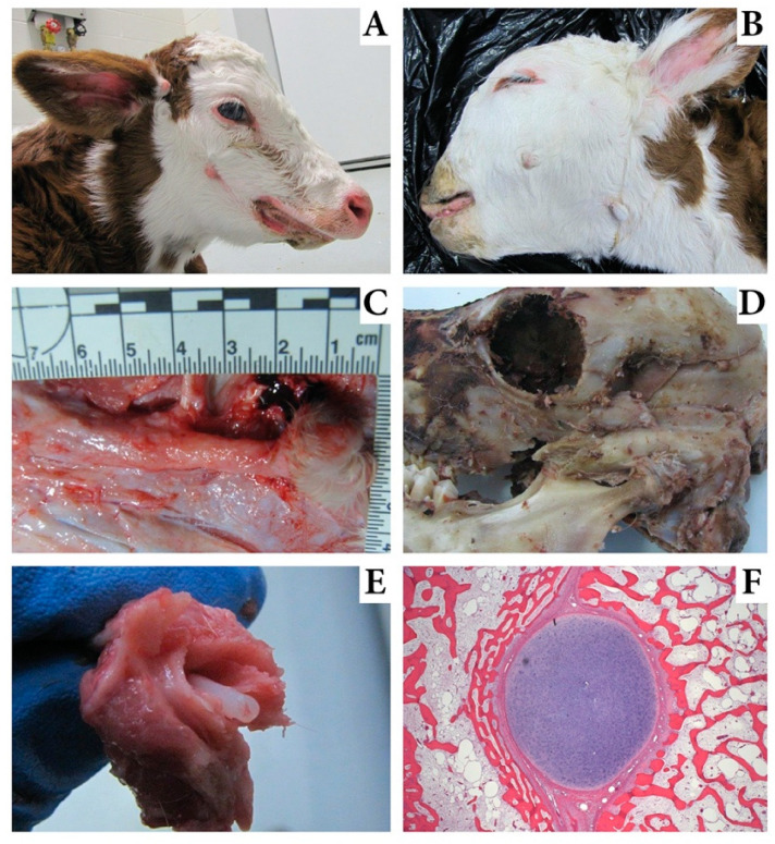
Figure 1.
Images of calves affected with mandibulofacial dysostosis (MD). (A). An MD calf with megastomia. Skin tags are visible ventral to the eye and at the base of the ear. Brachygnathia is also evident and a slight facial bulge is seen dorsal and caudal to the skin tag. (B). An MD calf with skin tags; one is caudal to commissure of the lips and one is ventral to the base of the ear near the caudal ramus of the mandible. (C). Exposure of the abnormal bone in an MD calf with the skin tag intact at the right margin. (D). The skull of an MD calf showing the exposed bone fractured during autopsy and demonstrating origin of this abnormal bone just above the temporal mandibular joint. (E). An image of the fractured bony prominence in an MD calf exposing the retained Meckel’s cartilage within the bony prominence. (F). Histology evaluation of the Meckel’s cartilage core from an MD calf surrounded by bone and separated by fibrous tissue.

