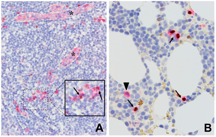
Figure 3.
Immunohistochemistry using antibodies against CD123 (red signal, surface/cytoplasmic) and TCF4 (brown signal, nuclear). (A) Normal tonsil showing CD123+/TCF4− expression on endothelial cells (asterisks), with a few scattered plasmacytoid dendritic cells showing CD123+/TCF4+ expression (arrow). (B) Bone marrow with scattered CD123+/TCF4− myeloid cells (arrowhead) as well as CD123+/TCF4+ plasmacytoid dendritic cells (arrows). (A and B. Main figures at 200× magnification (inset, 400×), hematoxylin counterstain).

