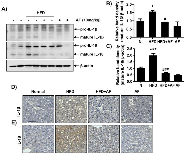
Figure 4.
Auranofin ameliorates interleukin (IL) 1-β and IL-18 expression and secretion in the liver and serum of the HFD-induced NAFLD mouse model. Following treatment with or without HFD and auranofin for 8 weeks, the liver tissues were obtained. (A) Liver (300 μg) was homogenized with 500 μL of lysis buffer supplemented with protease and phosphatase inhibitors. Western blots showing levels of IL-1β and IL-18 in the mouse liver in each group. Quantification of IL-1β (B) and IL-18 (C) expression. Data are expressed as means ± SD (n = 4). * p < 0.05 and *** p < 0.001 compared with the normal group. # p < 0.05 and ### p < 0.001 compared with HFD group. The hepatic expression of IL-1β (D) and IL-18 (E) protein were assessed by immunohistochemistry analysis (n = 3). Scale bar—50 μm. N, normal group; HFD, HFD fed group; HFD+AF, HFD fed and auranofin injection group; AF, auranofin injection group.

