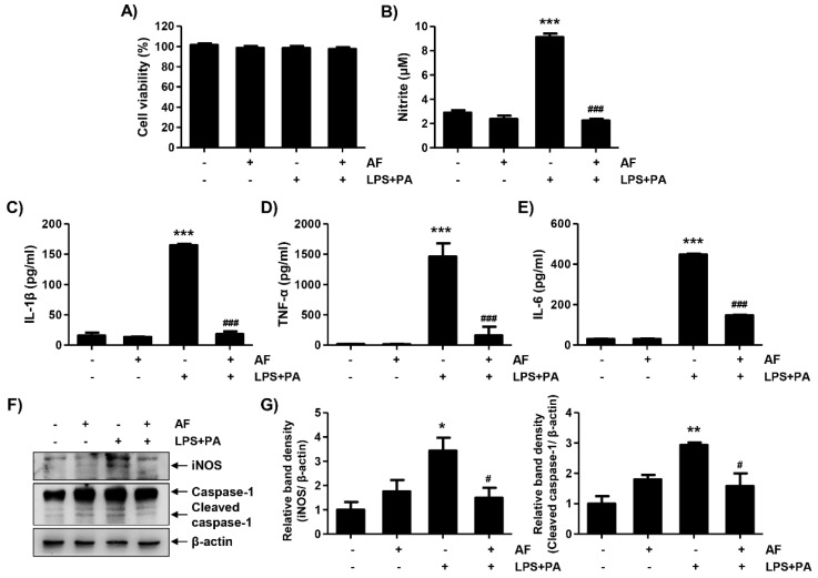
Figure 7.
Auranofin suppresses LPS and PA-induced inflammation in primary hepatocytes. The cells were pretreated with auranofin (1.5 μM) for 1 h and treated with LPS and PA for 24 h. (A) Cell viability was estimated by 3-(4,5-dimethylthiazol-2-yl)-2,5-diphenyltetrazolium bromide (MTT) assay. The absorbance was measured using the microplate reader at 540 nm and compared to the control which was set to 100%. Data are expressed as means ± SD (n = 6). (B–E) Nitrite and proinflammatory cytokines, including IL-1β, TNF-α and IL-6, were assessed using Griess reagent and ELISA kit. The absorbance was measured using the microplate reader. The error bars represent the standard deviation of three independent experiments. Statistical analysis was performed using an ANOVA with Tukey’s post-hoc test (n = 6). (F) The cells were lysed, and a Western blot was performed to evaluate the expression of inducible nitric oxide synthase and caspase-1. Equal protein loading was confirmed by β-actin expression. (G) Quantification of iNOS and activated caspase-1 expression. Data are expressed as means ± SD (n = 2). * p < 0.05, ** p < 0.01, and *** p < 0.001 compared with un-treated cells. # p < 0.05 and ### p < 0.001 compared with LPS and PA-treated cells. AF, auranofin; PA, palmitic acid; LPS, lipopolysaccharide.

