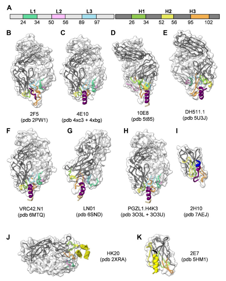
Figure 2.
Structures of gp41-specific antibodies in complex with their epitope. The Fabs are represented in ribbon colored according to the light and heavy chain scheme, highlighting the position of the different CDRs (A). The surface of the Fabs is represented by a semi-transparent white surface. The gp41 epitope is represented as cartoon colored according to the gp41 scheme shown in Figure 1A. The PBD coordinate files used for generating the figure for each antibody are indicated. (B) 2F5; (C) 4E10; (D) 10E8; (E) DH511.1; (F) VRC42.N1; (G) LN01; (H) PGZL1.H4K3; (I) llama nanobody 2H10; (J) HK20 targeting HR1; (K) llama nanobody 2E7 targeting HR1-CC. Images shown in Figure 2, Figure 3 and Figure 4 were rendered by ChimeraX [86].

