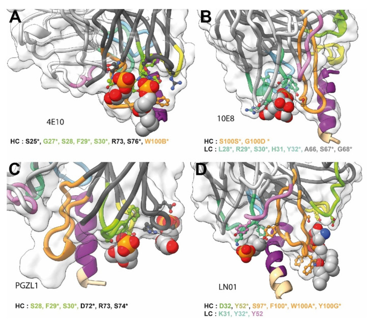
Figure 5.
Close-ups of lipid or lipid fragment binding of MPER bnAbs. (A) 4E10; (B) 10E8; (C) LN01 and (D) PGZL1. The gp41 epitope and Fabs are represented as in Figure 2. The lipid or lipid fragment visible in the structures are shown as spheres. The residues coordinating the binding of the lipids are shown as stick and balls, the identity and number of these residues are indicated below each structure, an asterisk indicates which residues are present in the UCA.

