
Figure 1.
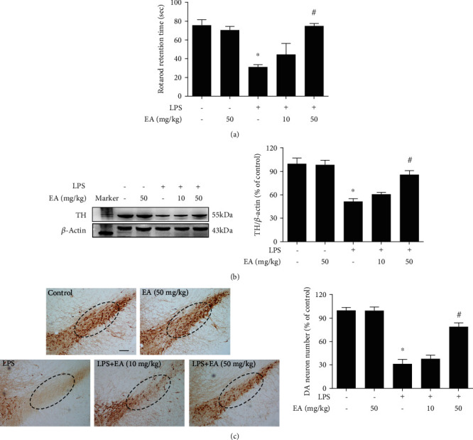
EA attenuated LPS-induced DA neuronal damage in SN in vivo. Rats were intragastrically given EA (50 mg/kg) for 7 consecutive days. Rat behavior changes were analyzed by the rotarod test (a). TH protein expression in the rat midbrain was tested by western blot assay (b). Brain sections were immunostained with an anti-TH antibody, and the number of TH-positive neurons in SN was counted (c). The “ellipse” presented the area of SN. Scalebar = 200μm. Data were the mean ± SEM from 6 rats. ∗p < 0.05 compared with the control group; #p < 0.05 compared with the LPS group.