
Figure 3.
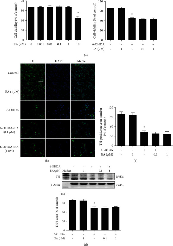
EA had no direct neuroprotective effects on DA neurons. MN9D cells were treated with EA (0.1 and 1 μM) for 30 min and then incubated with 6-OHDA (100 μM) for 24 h. Cell viability was determined by MTT assay (a). 6-OHDA-induced MN9D cell damage was evaluated by immunostaining (b) and cell counting (c). Scalebar = 100μm. The protein expression of TH was detected by western blot assay (c). Data were the mean ± SEM from three independent experiments performed in triplicate. ∗p < 0.05 compared with control cultures; #p < 0.05 compared with 6-OHDA-treated cultures.