
Figure 1.
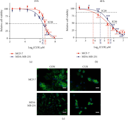
Breast cancer cell viability is inhibited by curcumin treatment. (a) Curcumin effectively inhibits the cell viability of cancer cells. Cancer cells were treated with the indicated concentrations of curcumin for 24 h. Cell viability was measured by the CCK-8 assay. Each point represents the mean value of three independent determinations; the error bars represent the SEM. ∗P < 0.05; ∗∗P < 0.01; ∗∗∗P < 0.001, compared to controls (MCF-7 cells). #P < 0.05; ##P < 0.01; ###P < 0.001, compared to controls (MDA-MB-231 cells). (b) Curcumin effectively inhibits the cell viability of cancer cells. Cancer cells were treated with the indicated concentrations of curcumin for 48 h. Cell viability was measured by the CCK-8 assay. Each point represents the mean value of three independent determinations; the error bars represent the SEM. ∗P < 0.05; ∗∗P < 0.01; ∗∗∗P < 0.001, compared to controls (MCF-7 cells). #P < 0.05; ##P < 0.01; ###P < 0.001, compared to controls (MDA-MB-231 cells). (c) MCF-7 cells were treated with 40 μM curcumin for 48 h (CUR), and MCF-MB-231 cells were treated with 50 μM curcumin for 48 h (CUR), with the control groups (CON) receiving the same volume of DMSO (curcumin solvent) as the treatment groups, and cell morphology was imaged using a fluorescence microscope (scale bar represents 100 μm). Representative images from three independent experiments are shown. All the CON groups treated above received the same volume of reagent solvent as the treatment groups.