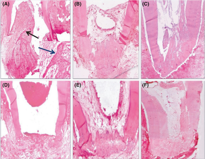
FIGURE 4.

(A) Photomicrograph of the apical third of a root in group I (blood clot) after 1 month showing differentiation of the epithelial cells (black arrow), no vital tissue infiltration, no sign of hard tissue formation, open apex and moderate inflammatory cell infiltration (H&E, ×40). Notice the well‐differentiated epithelial cells migrating towards the root apex (blue arrow) as seen in the small box (H&E, ×200). (B) Photomicrograph of the apical third of a root in group I after 2 months showing vital tissue infiltration towards the apical third, partial hard tissue formation, signs of apical closure and mild inflammatory cell infiltration (H&E, ×40). (C) Photomicrograph of the apical two‐thirds of a root in group I after 3 months showing vital tissue infiltration along the apical two‐thirds of the pulp space, fibrous tissue formation and calcified structures (black arrow) inside the pulp space, partial hard tissue formation, signs of apical closure and mild inflammation (H&E, ×40). (D) Photomicrograph of the apical third of a root in group II (17% EDTA + blood clot) after 1 month showing no vital tissue infiltration, signs of partial hard tissue formation, open apex and mild inflammation (H&E, ×40). (E) Photomicrograph of the apical third of a root in group II after 2 months showing vital tissue infiltration at the apical third, partial hard tissue formation, signs of apical closure and mild inflammation (H&E, ×40). (F) Photomicrograph of the apical two‐thirds of a root in group II after 3 months showing vital tissue infiltration along the pulp space, fibrous tissue formation inside the pulp space, signs of complete hard tissue formation, apical closure and mild inflammation (H&E, ×40)
