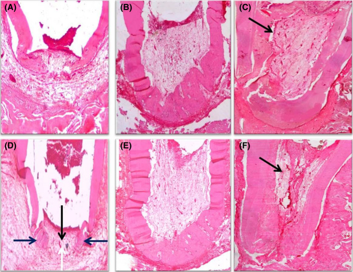
FIGURE 5.

(A) Photomicrograph of the apical third of a root in group III (PRF) after 1 month showing vital tissue infiltration into the apical third, signs of partial hard tissue formation, open apex, mild inflammation and a line of demarcation separating the newly formed hard tissue from the old one (black arrow) (H&E, ×40). (B) Photomicrograph of the apical half of a root in group III after 2 months showing vital tissue infiltration at the apical third, partial hard tissue formation, signs of apical closure and no sign of inflammation (H&E, ×40). (C) Photomicrograph of the apical two‐thirds of a root in group III after 3 months showing vital tissue infiltration along the pulp space, odontoblast‐like cells (black arrow), increased vascularity inside the pulp space, complete hard tissue formation, signs of apical closure and no inflammation (H&E, ×40). (D) Photomicrograph of the apical third of a root in group VI (17% EDTA + PRF) after 1 month showing vital tissue infiltration at the apical third (black arrow), signs of partial hard tissue formation (blue arrows), open apex (white arrow) and mild inflammation (H&E, ×40). (E) Photomicrograph of the apical half of a root in group IV after 2 months showing vital tissue infiltration towards the middle third, partial hard tissue formation, signs of apical closure and no sign of inflammation (H&E, ×40). (F) Photomicrograph of the apical two‐thirds of a root in group IV after 3 months showing vital tissue infiltration along the pulp space, odontoblast‐like cells undergoing differentiation (black arrow), increased vascularity inside the pulp space, complete hard tissue formation, signs of apical closure and no sign of inflammation (H&E, ×40)
