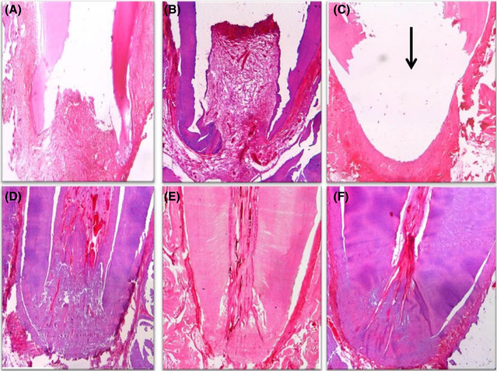
FIGURE 6.

(A) Photomicrograph of the apical third of a root in group V (positive control) after 1 month showing no vital tissue infiltration, no signs of hard tissue formation, open apex, severe inflammation and granulation tissue formation (H&E, ×40). (B) Photomicrograph of the apical half of a root in group V after 2 months showing the same findings after 1 month with increased granulation tissue (H&E, ×40). (C) Photomicrograph of the apical third of a root in group V after 3 months showing the same findings after 1 month with abscess cavity formation (black arrow) (H&E, ×40). Photomicrographs of the apical third of a root in group VI (negative control) after 1 month (D), 2 months (E) and 3 months (F) showing normal architecture of pulpal tissue, signs of apical closure and no inflammation (H&E, ×40)
