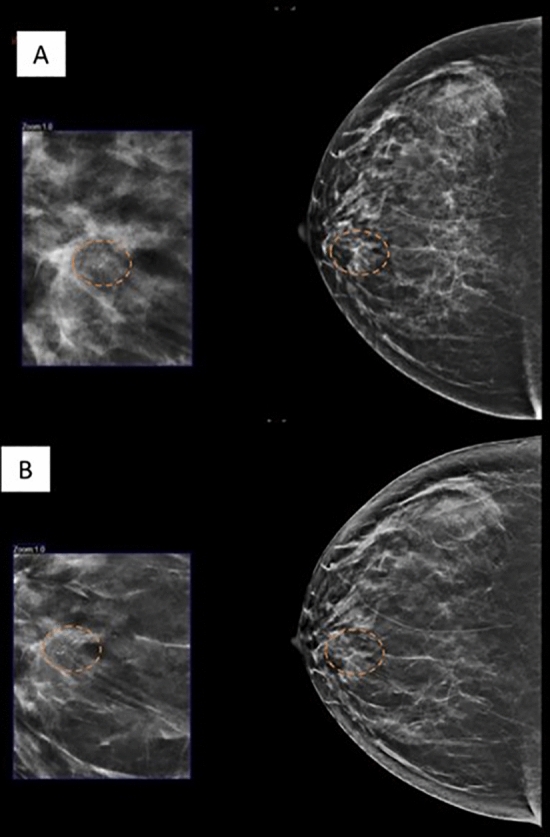
Figure 5.

46 year-old lady who came for screening mammography. CC view of the right breast in FFDM (A) and C-view (C-view software by Hologic, software version 1.7, http://www.lowdose3d.com/) (B) showing a suspicious calcification in linear distribution in the inner quadrant (dashed circle). Vacuum assisted stereotactic guided biopsy was performed. The distribution of the calcifications were clearly seen in the synthesized 2D image from tomosynthesis. HPE proven DCIS.
