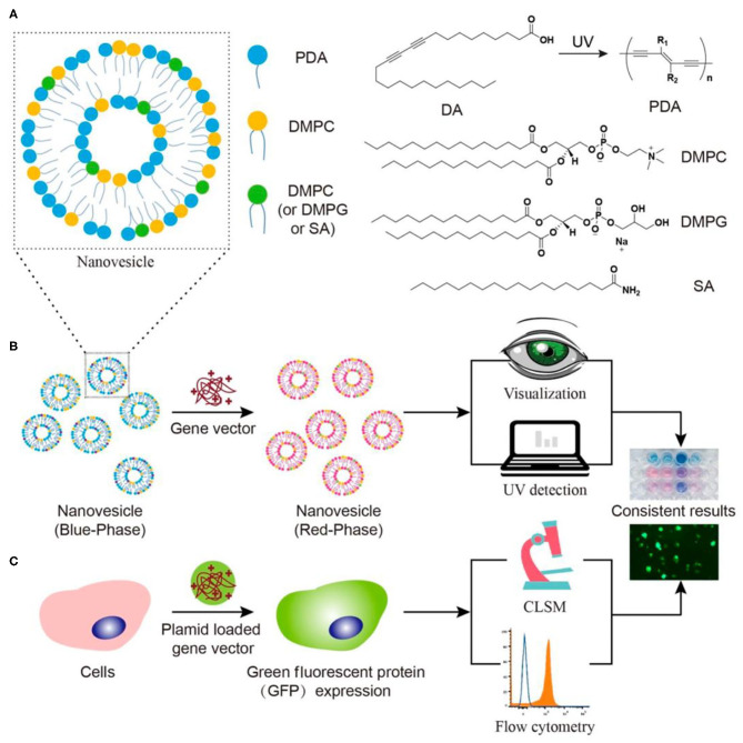
Figure 9.
Schematic illustration of (A) the structure of nanovesicle composed of DA, PDA, DMPC, and/or DMPG/SA. (B) Fast visualization detection of the membrane affinity of gene vectors using PDA nanovesicles, compared to (C) a traditional cell transfection method. Reprinted with permission from Wang et al. (2019a). Copyright 2019, American Chemical Society.

